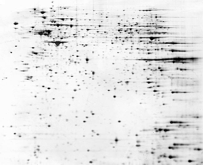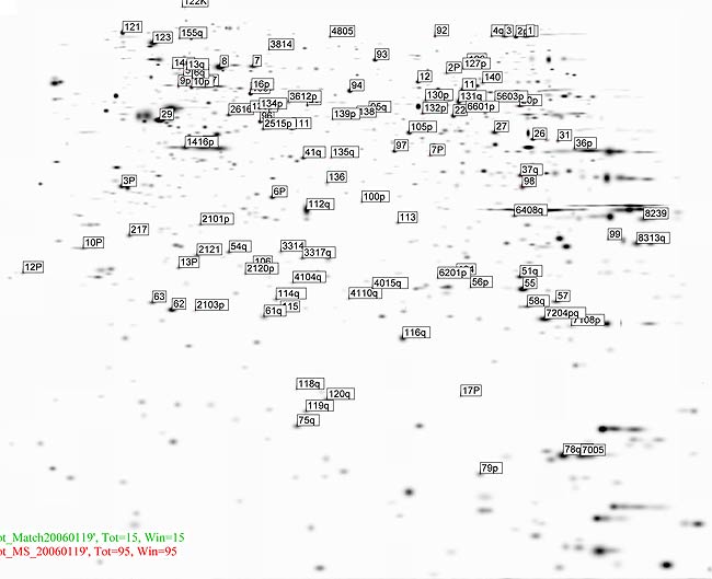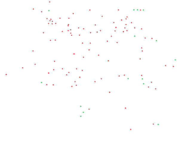
| Interactive 2-DE gel map of total extract of anti-IgM stimulated Ramos B cells. The Proteins are separated in a nonlinear pH gradient 3-10 and detected by silver staining. |
| Proteins highlighted in red are identified by peptide mass fingerprinting (PMF) and/or by fragment ion analysis. Proteins highlighted in green are identified by comparing their positions to the known proteins in other lymphocyte 2-DE databases. Move the mouse pointer over a protein spot to see the UniProt entry name of the protein and the spot number referring to Tables 1, 3-4 and Supplementary Tables 1-2. Click on the spot to get UniProt information about the protein. |


|
|||||||||||||||||||||||||||||||||||